


8 th Musculoskeletal MRI Online Reporting Workshop
Master Course in Fetal Radiology
8th Musculoskeletal MRI Online Reporting Workshop
Master Course in Fetal Radiology
8th Musculoskeletal MRI Online Reporting Workshop
Master Course in Fetal Radiology
A Hybrid Obstetric Ultrasound Training Course for
Radiologists and Residents
.
DEPARTMENT OF RADIODIAGNOSIS
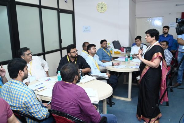
Event Overview
Moderator
Expert Panel
Presenting Doctors
The supporting faculty contributed their expertise to the discussions, assisting in the case- based learning and providing clinical perspectives to the participants.
Webinar Description
This webinar focused on the evaluation of adnexal masses using ultrasound, a critical aspect of gynecological diagnostics and patient management. The format included:
Learning Objectives
• Understand sonographic features of adnexal masses.
• Learn practical application of diagnostic criteria in real-world scenarios.
• Gain expert insight into differentiating benign and malignant lesions.
• Promote critical thinking and collaborative case analysis.
DEPARTMENT OF RADIODIAGNOSIS
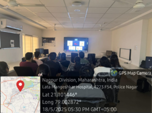
Title: Master Course in Fetal Radiology
Organized by: Department of Radio-diagnosis, NKPSIMS
Date: 1st June 2025
Time: 9:00 AM – 2:00 PM
Venue: Department of Radio-diagnosis, NKPSIMS
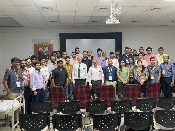
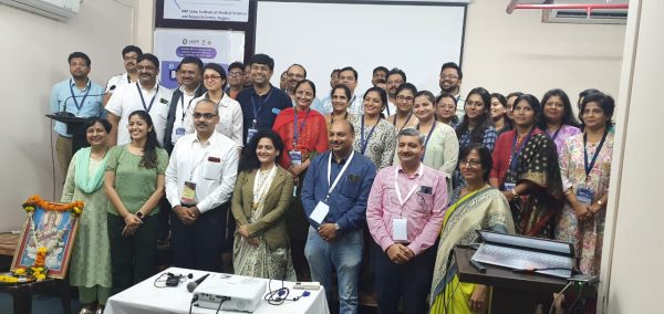
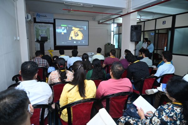
The Department of Radio-diagnosis, NKPSIMS, successfully organized the Master Course in Fetal Radiology on 1st June 2025, from 9:00 AM to 2:00 PM. The course focused on enhancing the diagnostic skills of clinicians and radiologists in interpreting complex antenatal (ANC) cases, with an emphasis on fetal radiology.
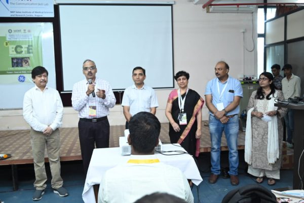
Lead Faculty:
Dr. Jyoti Chaubal
Dr. Alpana Joshi
Dr. Shilpa Sartarkar
Dr. Sunil Kabra
These renowned experts in fetal medicine delivered interactive and comprehensive lectures on a series of special ANC cases, offering valuable insights into prenatal imaging techniques and the interpretation of complex fetal anomalies. Their depth of knowledge and teaching methods made the sessions both engaging and highly informative.
Supporting Faculty:
Dr. Prashant Onkar
Dr. Dipali Kadam
Dr. Sandeep Dhote
Dr. Jitendra Sahu
The supporting faculty contributed their expertise to the discussions, assisting in the case-based learning and providing clinical perspectives to the participants.
Course Coordinator:
Dr. Prashant Onkar
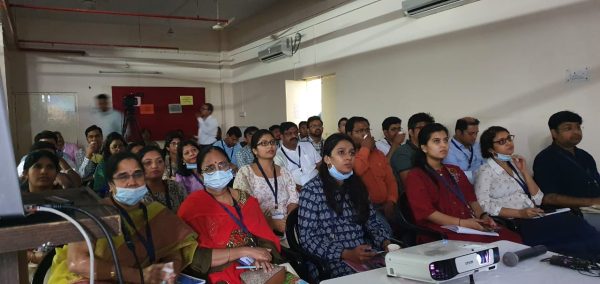
The event was well-attended by postgraduate students, radiologists, and clinicians, who actively participated in discussions and case analyses. The feedback received was highly positive, with participants highlighting the educational value and clinical relevance of the sessions. This course is part of the department’s ongoing efforts to provide advanced training in fetal radiology and to promote high standards in prenatal diagnostics.
DEPARTMENT OF RADIODIAGNOSIS
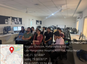
Title: Master Course in Fetal Radiology
Organized by: Department of Radio-diagnosis, NKPSIMS
Date: 1st June 2025
Time: 9:00 AM – 2:00 PM
Venue: Department of Radio-diagnosis, NKPSIMS
The Department of Radio-diagnosis, NKPSIMS, successfully organized the Master Course in Fetal Radiology on 1st June 2025, from 9:00 AM to 2:00 PM. The course focused on enhancing the diagnostic skills of clinicians and radiologists in interpreting complex antenatal (ANC) cases, with an emphasis on fetal radiology.
Lead Faculty:
Dr. Jyoti Chaubal
Dr. Alpana Joshi
Dr. Shilpa Sartarkar
Dr. Sunil Kabra
These renowned experts in fetal medicine delivered interactive and comprehensive lectures on a series of special ANC cases, offering valuable insights into prenatal imaging techniques and the interpretation of complex fetal anomalies. Their depth of knowledge and teaching methods made the sessions both engaging and highly informative.
Supporting Faculty:
Dr. Prashant Onkar
Dr. Dipali Kadam
Dr. Sandeep Dhote
Dr. Jitendra Sahu
The supporting faculty contributed their expertise to the discussions, assisting in the case-based learning and providing clinical perspectives to the participants.
Course Coordinator:
Dr. Prashant Onkar
The event was well-attended by postgraduate students, radiologists, and clinicians, who actively participated in discussions and case analyses. The feedback received was highly positive, with participants highlighting the educational value and clinical relevance of the sessions. This course is part of the department’s ongoing efforts to provide advanced training in fetal radiology and to promote high standards in prenatal diagnostics.
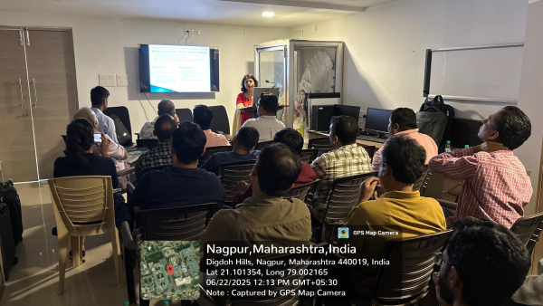
Event Overview
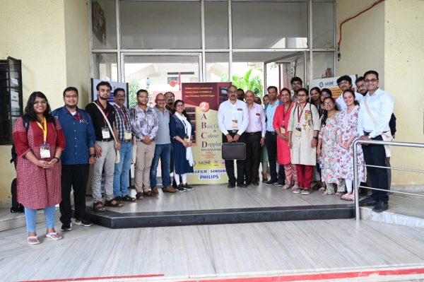
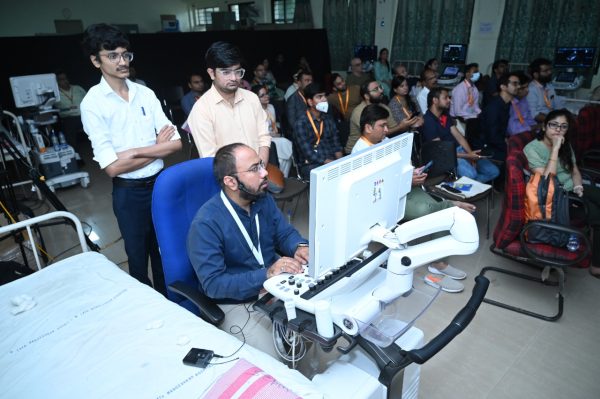
• Title: Level-II Course in Colour Doppler
• Organizer: Radiology Skill Academy Nagpur (RSAN)
•An initiative by the Department of Radiodiagnosis,
NKPSIMS
•Research Centre and LMH, Nagpur
• Venue : NKPSIMS & RC and LMH, Hingna Road, Nagpur
Duration & Dates:
• Four Days On-Site Training
• Dates: 19th to 22nd June 2025 (Thursday to Sunday)
• Timing: 9:00 AM to 5:00 PM
Target Audience:
• Radiologists
• Radiology Residents
Course Coordinators:
• Dr. Varsha Rangankar – Professor, Department of Radiodiagnosis, DY
Patil Medical College
• Dr. Prashant Onkar – Professor, Department of Radiodiagnosis, NKPSIMS
& RC and LMH, Nagpur
Course highlights:
• Intensive hands-on training in Colour Doppler techniques
• Focused on "Do it Yourself" approach
• Pre and Post tests (compulsory)
• Standard image storing exercise for each participant
• Image review and certification
• Access to supporting literature and videos (online)
• Post-course doubt solving sessions
Topics Covered:
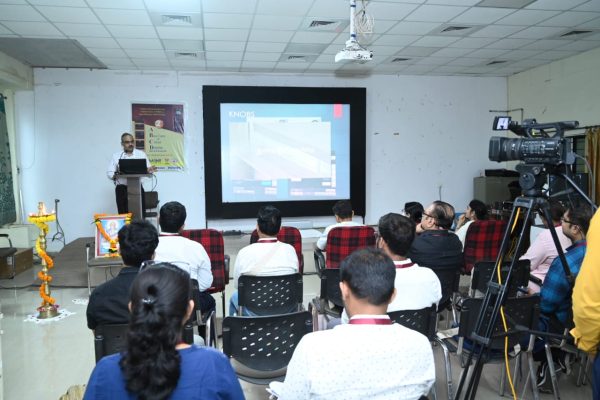
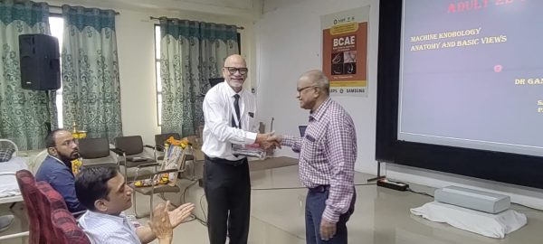
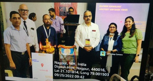
1. Knobology & Machine Settings
2. Carotid Doppler
3. Renal Doppler
4. Portal Doppler
5. Upper Limb Arterial & Venous
6. Lower Limb Arterial & Venous
7. Scrotal Doppler
8. Penile Doppler
Learning Objectives
• Understand sonographic features .
• Learn practical application of diagnostic criteria in real-world scenarios.
• Promote critical thinking and collaborative case analysis.
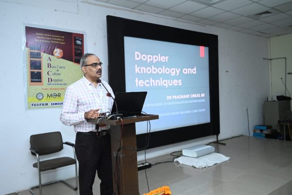
Colour Doppler studies are used along with ultrasound and have a longer learning curve than ultrasound. Due to multiple technical factors the students and junior radiologists find it difficult to master. There are no hands-on training programs available exclusively for colour Doppler. We have devised two courses for radiologists who want to master Doppler.
1. The basic course is for two days and covers all areas of peripheral doppler including carotid, upper and lower limb arterial and venous, renal and portal doppler. It is conducted in small batches with a teacher to student ratio 1:10. There are live demonstrations, hands- on experience and case discussions.
It is aimed for beginners as well as practitioners who do not have musc prior experience.
The level II course for four days consists of theory as well as practicals. It covers all aspects of color Doppler in detail. It includes live demonstrations, hand on experience and case discussions. In addition it also requires the participant to learn accurate image acquisitions. The images are evaluated by the faculty before awarding the certificate.
We have trained more than 150 radiologists in colour doppler in two years.Director – Dr Prashant Onkar, Professor.
For getting details of future batches please click on the following tab…
Echocardiography is actually ultrasound of the heart. Radiologists are well versed with anatomy of the heart and techniques of ultrasound. In many places there is dearth of physicians or cardiologists and therefore for getting echo studies done for pre surgical fitness or health check ups patients have to travel long distances. Though covered in the post-graduate radiology curriculum, it is not taught at many centers. We have devised a Basic course in Adult Echo.
It’s a two-day program with a very limited batch of participants. there are demonstrations of techniques, lectures and hands-on experience for all participants and case-based discussions.
For getting details of future batches please click on the following tab…
.
Magnetic resonance imaging has changed the detection of diseases significantly due to its advantages. There has been continuous development in the field of MRI in terms of machine capacity, imaging sequences, protocols etc. The advent of 3 Tesla MRI equipment has increased its use in musculoskeletal diseases and usually the final investigation performed for their evaluation. The information available from MRI studies is enormous and needs detailed knowledge of anatomy and pathology. MSK radiology has come up as a sub-specialty in recent decades.
We have devised this online course with an aim
The highlights are
For getting details of future batches please click on the following tab..
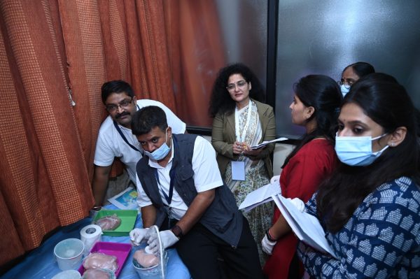
All radiologists are entitled to perform fetal interventions. Various newer techniques are now available for prenatal diagnosis of genetic diseases. They require sampling of amniotic fluid, chorion etc. Other interventions like blood transfusion, cord sampling are also on the rise. There is an increased need for skilled people who can perform fetal interventions. A radiologist is well versed with image guided interventional techniques like drainage FNA and Biopsy. Considering this gap we have designed a few models for the training in basic techniques of fetal interventions. We use them to train the radiologist during the Basic course in fetal interventions
Dr Jitendra Sahu, fetal radiologist is conducting these courses.
For getting details of future batches please click on the following tab…
.
.
.
Colour Doppler studies are used along with ultrasound and have a longer learning curve than ultrasound. Due to multiple technical factors the students and junior radiologists find it difficult to master. There are no hands-on training programs available exclusively for colour Doppler. We have devised two courses for radiologists who want to master Doppler.
1. The basic course is for two days and covers all areas of peripheral doppler including carotid, upper and lower limb arterial and venous, renal and portal doppler. It is conducted in small batches with a teacher to student ratio 1:10. There are live demonstrations, hands- on experience and case discussions.
It is aimed for beginners as well as practitioners who do not have musc prior experience.
The level II course for four days consists of theory as well as practicals. It covers all aspects of color Doppler in detail. It includes live demonstrations, hand on experience and case discussions. In addition it also requires the participant to learn accurate image acquisitions. The images are evaluated by the faculty before awarding the certificate.
We have trained more than 150 radiologists in colour doppler in two years.Director – Dr Prashant Onkar, Professor.
For getting details of future batches please click on the following tab…
Echocardiography is actually ultrasound of the heart. Radiologists are well versed with anatomy of the heart and techniques of ultrasound. In many places there is dearth of physicians or cardiologists and therefore for getting echo studies done for pre surgical fitness or health check ups patients have to travel long distances. Though covered in the post-graduate radiology curriculum, it is not taught at many centers. We have devised a Basic course in Adult Echo.
It’s a two-day program with a very limited batch of participants. there are demonstrations of techniques, lectures and hands-on experience for all participants and case-based discussions.
For getting details of future batches please click on the following tab…
.
Magnetic resonance imaging has changed the detection of diseases significantly due to its advantages. There has been continuous development in the field of MRI in terms of machine capacity, imaging sequences, protocols etc. The advent of 3 Tesla MRI equipment has increased its use in musculoskeletal diseases and usually the final investigation performed for their evaluation. The information available from MRI studies is enormous and needs detailed knowledge of anatomy and pathology. MSK radiology has come up as a sub-specialty in recent decades.
We have devised this online course with an aim
The highlights are
For getting details of future batches please click on the following tab..
All radiologists are entitled to perform fetal interventions. Various newer techniques are now available for prenatal diagnosis of genetic diseases. They require sampling of amniotic fluid, chorion etc. Other interventions like blood transfusion, cord sampling are also on the rise. There is an increased need for skilled people who can perform fetal interventions. A radiologist is well versed with image guided interventional techniques like drainage FNA and Biopsy. Considering this gap we have designed a few models for the training in basic techniques of fetal interventions. We use them to train the radiologist during the Basic course in fetal interventions
Dr Jitendra Sahu, fetal radiologist is conducting these courses.
For getting details of future batches please click on the following tab…